Products
3D Similution & 3D-printed Products
The application of personalized 3D printing technology in medicine is making significant strides in the development of implant devices, particularly artificial bones and joints. These 3D-printed implants are designed entirely based on the anatomical structure of each patient, utilizing data from CT and MRI scans. This approach ensures the creation of highly accurate devices that fit perfectly with the patient’s body, minimizing the risk of errors or incompatibility often seen with traditional implants.
Key Features:
- Complete Personalization: Each implant is designed and 3D-printed based on the specific physical characteristics and medical condition of the patient. This ensures that the device is highly compatible with the individual’s bone and joint structure, optimizing stability and function after surgery.
- Biocompatible Titanium Alloy Material: The artificial bones and joints are printed using durable biocompatible materials that are safe for the body and can naturally integrate with bone tissue.
- Minimized Postoperative Complications: Thanks to precise design and high compatibility, 3D-printed implants help reduce common postoperative issues such as infections, rejection, and other complications.
- Rapid Production Process: 3D printing technology shortens the production time of implants, from design to the final product, allowing patients to undergo surgery and begin recovery more quickly. This is especially crucial for cancer patients, where treatment time is of utmost importance.
- Versatile Applications: Personalized 3D-printed implants can be used for various parts of the body, from the spine and pelvis to knee and hip joints, ensuring suitability for complex orthopedic reconstructive surgeries.
With advanced technology and high precision, personalized 3D-printed implants not only offer superior treatment outcomes but also enhance quality of life for patients, helping them quickly return to daily activities with confidence and comfort.
If you have any questions or need further information, please contact us at:
3D TECHNOLOGY IN MEDICINE CENTER
Address: G-building, VinUniversity, Da Ton, Gia Lam, Hanoi
Phone: 0374 98 1111 (Dr. Hoang)
Email: 3dcenter@vinuni.edu.vn
Fanpage: https://www.facebook.com/vin3dlab
Youtube: https://www.youtube.com/@3DLABVINUNI


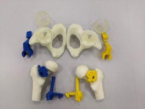
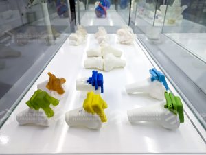
Patient-specific instrumentation (PSI) is an advanced solution that provides maximum support for complex surgeries, ensuring high precision and optimal outcomes. PSI is custom-designed based on the specific anatomical structure of each patient, using modern 3D printing technology and medical imaging data from CT and MRI scans. This allows surgeons to perform procedures accurately and swiftly, particularly in orthopedic surgeries, joint replacements, or spinal surgeries.
Key Features:
- Patient-Specific Design: Each PSI is 3D-printed based on the patient’s unique anatomical model, providing precise guidance for bone cutting, position adjustments, or joint replacements. This ensures that surgical steps are executed as planned, minimizing deviations during the procedure.
- High Precision: With personalized design and advanced 3D printing technology, PSI helps surgeons accurately position instruments and devices, reducing reliance on conventional navigation tools. This is especially crucial in bone and spinal surgeries, where even minor deviations can lead to serious issues.
- Reduced Surgery Time: PSI optimizes the surgical process, reducing the overall duration of the procedure. This not only lessens the surgeon’s stress but also minimizes the risk of infection and postoperative complications for the patient.
- Safe and Highly Effective: Using PSI improves surgical outcomes and reduces complications such as infections, blood loss, or damage to surrounding tissues. With precise planning and execution, PSI enables faster recovery and shorter hospital stays for patients.
- Versatile Applications: PSI can be used in a wide range of surgeries, including knee and hip replacements, spinal surgeries, bone realignment, and other complex procedures. It is particularly useful in cases requiring absolute precision for complex conditions.
PSI Development Process:
The development of PSI begins by gathering the patient’s medical imaging data through CT or MRI scans. These data are then used to create a 3D model of the surgical area. Based on this model, biomedical engineers design a unique 3D-printed navigation device that matches the patient’s specific anatomy. Once the design is finalized, the PSI is 3D-printed with high precision, ready to assist the surgeon during the operation.
Patient-specific instrumentation (PSI) represents a major advancement in medicine and a vital tool for enhancing safety, accuracy, and efficiency in surgery. With the support of PSI, surgical procedures carry less risk, yield better results for patients, and help them recover quickly after treatment.
Simulation of Using Patient-specific instrumentation (PSI) in Total Hip Replacement Surgery
Simulation of Using Patient-specific instrumentation (PSI) in the Correction of Multidirectional Deformed Bone Alignment
If you have any questions or need further information, please contact us at:
3D TECHNOLOGY IN MEDICINE CENTER
Address: G-building, VinUniversity, Da Ton, Gia Lam, Hanoi
Phone: 0374 98 1111 (Dr. Hoang)
Email: 3dcenter@vinuni.edu.vn
Fanpage: https://www.facebook.com/vin3dlab
Youtube: https://www.youtube.com/@3DLABVINUNI
In the digital age, Digital 3D Planning offers an advanced, flexible, and cost-effective solution for design and surgical processes. Without the need to produce physical models, digital 3D plans allow doctors, engineers, and medical specialists to directly view, manipulate, and plan on 3D models via digital devices. This optimizes the surgical preparation process and enhances collaboration between medical experts worldwide, remotely.
Key Features:
- No Need for Physical Models: Instead of creating a physical 3D-printed model, digital 3D plans allow the use of models directly on computer or mobile screens. This saves costs, reduces waiting times, and enables easy, immediate adjustments or modifications.
- Personalized Customization: The 3D plan is built based on each patient’s medical data, accurately simulating their specific anatomy and medical condition. Specialists can easily analyze and adjust the surgical plan for individual cases without the need for complex production steps.
- Integration of Virtual Reality and Augmented Reality (VR/AR): Digital 3D plans can integrate with VR and AR technologies, providing surgeons with a more intuitive experience when reviewing the patient’s anatomical details. This improves accuracy and enhances surgical preparation.
- Easy Remote Interaction: Digital 3D plans can be easily shared over the internet, allowing medical professionals from various locations to discuss and create treatment plans together. This is a powerful tool for international collaboration, especially in complex cases that require input from top specialists.
- Manipulation and Viewing from All Angles: With digital 3D plans, doctors can rotate, zoom in, zoom out, and inspect every angle of the model, providing them with both an overall and detailed view of the upcoming surgery.
- Environmentally Friendly: Using digital plans eliminates the need for printing materials, contributing to reduced environmental impact.
Medical Applications:
Digital 3D planning has been widely applied across various medical fields, from orthopedic surgery, dentistry, and neurology to complex cosmetic surgeries. With its optimal customization capabilities, these plans allow doctors to prepare thoroughly, minimize surgical risks, and ensure the best possible outcomes for patients.
Benefits in Time and Cost
Fully digitizing the planning process not only reduces the waiting time for producing physical products but also saves costs associated with materials, 3D printers, and equipment maintenance. This is particularly crucial in situations that require speed and flexibility, such as emergencies or urgent surgeries.
Digital 3D Planning represents a new advancement in modern medicine, enhancing the quality of patient care and opening up new opportunities for applying digital technology in medical procedures. Due to its flexibility and efficiency, this solution will continue to develop and become an indispensable tool in future surgical processes.
If you have any questions or need further information, please contact us at:
3D TECHNOLOGY IN MEDICINE CENTER
Address: G-building, VinUniversity, Da Ton, Gia Lam, Hanoi
Phone: 0374 98 1111 (Dr. Hoang)
Email: 3dcenter@vinuni.edu.vn
Fanpage: https://www.facebook.com/vin3dlab
Youtube: https://www.youtube.com/@3DLABVINUNI

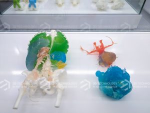
The Personalized 3D-Printed Anatomical Model is marking a turning point in modern medicine, providing more accurate surgical preparation and planning than ever before. Based on medical imaging data from methods such as CT scans and MRIs, these 3D anatomical models are custom-designed to match each patient’s body structure. This technology not only supports doctors in surgical planning but also offers enhanced opportunities for advanced training and research in the medical field.
Key Features:
- Fully personalized design: Each 3D anatomical model is printed according to the specific anatomical structure of the individual patient, precisely recreating details such as bones, blood vessels, and soft tissues. This allows doctors to examine and analyze the case in detail before surgery, particularly beneficial for complex procedures.
- Surgical planning support: The 3D model allows doctors to simulate the entire surgical process before operating on the real body, optimizing the approach, minimizing risks, and ensuring successful outcomes. This tool is especially important in surgeries such as joint replacements, tumor removal, or bone reconstruction.
- Training and teaching: The 3D-printed anatomical model not only plays a role in clinical practice but also serves as an excellent teaching tool for medical students and doctors in training. Instead of relying solely on textbooks or generic models, they can learn from realistic anatomical models that accurately replicate human body structures.
- High precision: Advanced 3D printing technology enables the accurate recreation of every anatomical detail, allowing doctors to capture every important feature of the patient’s body, reducing errors during surgery.
- Minimizing complications: With thorough preparation using a 3D model, doctors can avoid unexpected risks during surgery, reducing post-operative complications and improving patient recovery.
Practical Applications:
- Orthopedics and trauma: 3D-printed models are widely used in orthopedic and trauma surgeries, helping simulate and prepare for joint replacements, bone splints, or bone reconstruction.
- Cardiovascular surgery: In surgeries related to blood vessels, the heart, and lungs, 3D models help doctors assess and plan the optimal intervention, minimizing risks during procedures.
- Neurosurgery: 3D anatomical models are also used in neurosurgery, allowing doctors to simulate complex areas of the brain and spine, better preparing for critical interventions.
- Dentistry: 3D-printed models are applied in dentistry for preparing dental implants, jawbone realignment, and other dental surgeries.
Model Development Process:
The development of a 3D-Printed Anatomical Model begins with collecting detailed medical imaging data of the patient through methods such as CT or MRI scans. Biomedical engineers then use specialized software to create a 3D model from this data, ensuring the model accurately reflects the patient’s body structure. Finally, the model is printed using advanced 3D printing technology, creating a detailed, accurate replica of the body part requiring surgery.
Benefits for Patients and Doctors:
- Improved treatment quality: The 3D-printed anatomical model helps doctors prepare better, thereby improving treatment outcomes and reducing risks for patients.
- Enhanced professional collaboration: These models can be shared among experts worldwide, allowing them to collaboratively evaluate and determine the optimal treatment for the patient.
- Reduced surgical time: With the 3D model, detailed surgical preparation can be conducted in advance, shortening surgery time and reducing the patient’s recovery period.
The Personalized 3D-Printed Anatomical Model has become a crucial tool in modern medicine, not only aiding doctors in preparing for complex surgeries but also supporting research and education. With its high precision and perfect customization, this technology opens new opportunities to enhance the quality of healthcare and patient treatment.
If you have any questions or need further information, please contact us at:
3D TECHNOLOGY IN MEDICINE CENTER
Address: G-building, VinUniversity, Da Ton, Gia Lam, Hanoi
Phone: 0374 98 1111 (Dr. Hoang)
Email: 3dcenter@vinuni.edu.vn
Fanpage: https://www.facebook.com/vin3dlab
Youtube: https://www.youtube.com/@3DLABVINUNI
Cranial-Brain Patch Model
In the field of neurosurgery, restoring and reconstructing the cranial structure after trauma or surgery is crucial. The Cranial-Brain Patch Model (also known as an artificial cranial-brain patch) is an advanced solution that replaces or repairs damaged areas of the skull, protecting the brain and restoring the natural anatomical shape of the cranium.
Key Features:
- Personalized Design: The cranial-brain patch model is precisely customized to each patient’s skull structure, based on medical imaging data such as CT or MRI scans. This ensures that the patch fits perfectly into the damaged area, providing stability and high aesthetic effectiveness.
- Advanced Materials: Cranial-brain patches are typically made from durable biocompatible materials such as titanium, PEEK (polyether ether ketone), or polymer-based biomaterials, ensuring strength, biocompatibility, and safety for the body. These materials also help reduce the risk of rejection and infection post-surgery.
- Functional and Aesthetic Restoration: The cranial-brain patch not only protects the brain from external impacts but also restores the natural shape of the skull, providing optimal aesthetic results, particularly in cases of severe damage or reconstruction surgeries.
- Excellent Bone Integration: The materials used for cranial-brain patches integrate well with natural bone tissue, supporting the healing process and creating a strong bond. This minimizes the risk of complications and ensures long-term effectiveness after surgery.
- 3D Printing Technology: Modern cranial-brain patches are often produced using 3D printing technology, offering high precision and customized solutions for each patient. This technology also reduces production time and minimizes errors during surgery.
Surgical Applications:
The cranial-brain patch model is commonly used in surgeries related to traumatic brain injuries, congenital skull defects, or cases where part of the skull needs to be removed due to tumors or other medical conditions. With personalized design and high compatibility, artificial cranial-brain patches help patients recover quickly and minimize post-surgery complications.
Production Process:
The production of cranial-brain patches begins by collecting patient imaging data through CT or MRI scans. A 3D model of the patient’s skull is created from this data, and biomedical engineers design the patch to fit the required size and shape. The patch is then 3D-printed from advanced biocompatible materials, ensuring high accuracy and readiness for surgery.
Benefits for Patients:
- Safety and Durability: Supported by modern technology and advanced materials, the cranial-brain patch provides a high level of safety and durability for patients.
- Optimized Recovery Process: Due to its high biocompatibility and regenerative properties, patients can recover quickly and return to normal life after surgery.
- Minimized Complications: The personalized design and high precision of the cranial-brain patch significantly reduce the risk of complications, such as infections, rejection, or displacement.
The Cranial-Brain Patch Model is an efficient and advanced solution in modern medicine, offering comprehensive recovery in both functional and aesthetic aspects for patients. The combination of 3D printing technology and advanced biomaterials is enhancing treatment quality in cranial surgeries, while improving patients’ overall quality of life.
If you have any questions or need further information, please contact us at:
3D TECHNOLOGY IN MEDICINE CENTER
Address: G-building, VinUniversity, Da Ton, Gia Lam, Hanoi
Phone: 0374 98 1111 (Dr. Hoang)
Email: 3dcenter@vinuni.edu.vn
Fanpage: https://www.facebook.com/vin3dlab
Youtube: https://www.youtube.com/@3DLABVINUNI

